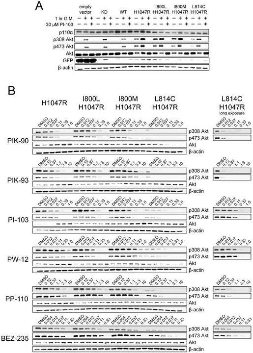
Figure 7. Yeast screen hits I800L, I800M, and L814C produce EGF-independent Akt phosphorylation in MCF10A cells, which maintains altered inhibitor sensitivities.

A: MCF10A cell lines expressing the indicated p110α mutants were cultured in growth media lacking EGF for 24 hours, and then treated with the indicated combinations of normal growth media (G. M.) and 30 μM PI-103. After 1 hour, the cells were lysed and subject to western blot analysis with the indicated antibodies.
B: The indicated MCF10A cell lines were cultured in growth media lacking EGF for 24 hours, and then treated for 1 hour with serial dilutions of the indicated PI3K inhibitors, after which the cells were lysed and subjected to western blot analysis with the indicated antibodies. The PI3K inhibitor concentrations are indicated in μM.
