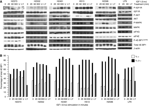
Figure 1.
IGF-I stimulation activates downstream-signalling pathways and inactivates 4E-BP1 in mesothelioma cell lines. (A) Immunoblot analysis showing phosphorylation and total levels of proteins involved in cell-signalling pathways (IGF1R, Akt, and MAPK) or in initiation of translation (eIF4G, eIF4E, and 4E-BP1) after treatment (in minutes) with and without IGF-I (5 nM) or on treatment with IGF-I (5 nM for 20 min) combined with LY249002 (LY) or U0126 (U). (B) Representation of the percentage of 4E-BP1 in hypophosphorylated isoforms (α, open columns) compared with that in hyperphosphorylated isoforms (β + γ, filled columns) for mesothelioma cells and mesothelial control cells (LP9) not treated or treated with IGF-I (5 nM) or IGF-I combined with LY249002 (LY) or U0126 (U) for the indicated times. β-actin represents loading controls for each cell line.

