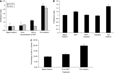
Figure 2.
Treatment with DHT results in an increase in cell proliferation in both normal and malignant ovarian cells. (A) OSEC2 and OVCAR 3 cells were stimulated for 24 h with DHT before analysis using an SRB assay. Both cell lines show increases in cell proliferation after DHT stimulation of up to 24 h. Increased cell proliferation is shown using a dose of 10 nM in the OSEC2 cells, but a dose of 100 nM in OVCAR3 cells. (B) OVCAR3 cells were treated for 24 h with 10 nM DHT in the presence and absence of the specific AR inhibitor, Casodex, before analysis using an SRB assay. The androgen-induced stimulation is abrogated by the addition of Casodex. Casodex alone had no effect on cell proliferation. (C) OVCAR3 cells were treated with DHT for 24 h and the S-phase fraction analysed using propidium iodide incorporation. Cells stimulated with DHT show a dose-dependent increase in the S-phase fraction compared with non-treated cells.

