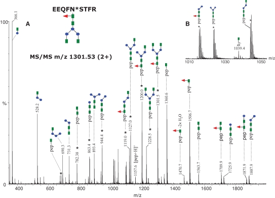
Fig. 2.
MS/MS of the precursor ion of m/z 1301.53 (2+) corresponding to the glycopeptide EEQFN*STFR, containing the glycan indicated at the top (left). Doubly charged ions are highlighted with an asterisk. The remaining ions are singly charged. (A) The MS/MS spectrum obtained in the data-dependent mode using a collision energy gradient from 30 to 40 V, showing all the observed fragment ions. (B) The inset in the mass range m/z 1010–1050 indicates the singly charged ion at m/z 1039.4 which arises from backbone cleavage and carries an N-acetylglucosamine residue. Color code: green square – N-acetylglucosamine; red triangle – fucose; blue circle – mannose; yellow circle– galactose; purple rhombus – sialic acid.

