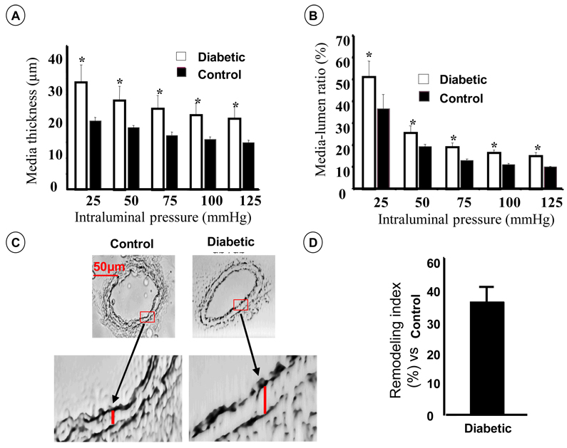
Figure 1.
Structural artery wall remodeling of mesenteric resistance artery from 12-week old type 2 diabetic and control mice. A: Media thickness measurements; B: Media/lumen ratio; C: Cross section of mesenteric resistance arteries from diabetic and control mice perfused in vivo with 4% of formalin, 60x magnification; D: Remodeling index (percentage of difference between internal diameters of diabetic and control vessels not attributable to growth), n=5–7; p<0.05 statistically different between diabetic vs. control. A–B. Resistance arteries were mounted in arteriograph for measurements of intraluminal and external diameter, and wall thickness under range of pressure from 25 to 125 mmHg.

