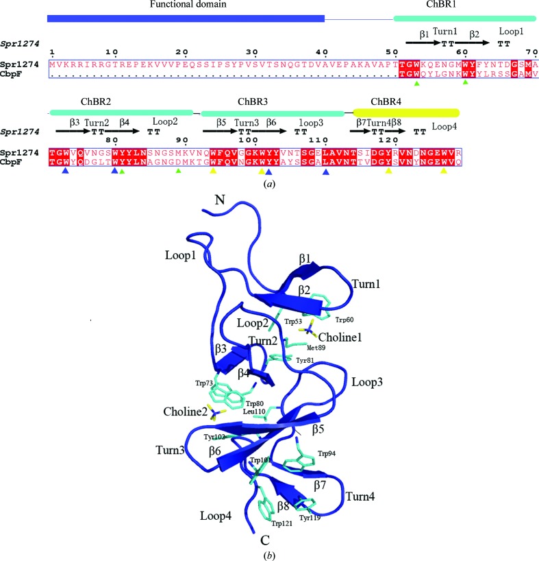
Figure 1.
(a) Sequence alignment of Spr1274 and the CBD of CbpF. The alignment was performed using ClustalX (Thompson et al., 1994 ▶) and ESPript (Gouet et al., 1999 ▶). The secondary-structure elements of Spr1274 are identified. The constituent domains of Spr1274 are displayed at the top of the alignment, with the functional domain in blue and the ChBRs in cyan or yellow (ChBR4 is not a canonical repeat). Residues involved in choline binding are indicated below with filled triangles (ChBS1, green; ChBS2, blue; ChBS3, yellow). (b) The overall structure of Spr1274-CBD (chain B). Secondary-structure elements are labelled (β-strands, turns and loops). The choline molecules (yellow) and residues (cyan) involved in choline binding are labelled and shown in stick representation.

