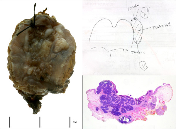
Figure 1.
Macroscopic view of laser resected tumour orientated by suture and clinical diagram. This shows the complexity of pathological interpretation which can be liable to sampling error. A whole mount view of H&E stained section of transverse slice through the tumour and tonsil show the close margin of excision. It is reasonable to assume that high quality 'real-time' pathological data would aid surgical incision and ensure a more complete excision. Retrospective analysis paraffin section H&E appears less useful since it cannot immediately inform surgery only later therapy. Optical diagnostics technology may provide a means to improve surgical treatment and eventual outcome by informing the surgeon in 'real-time' and improving the margin; (Courtesy of Dr A Sandison, Imperial College, London).

