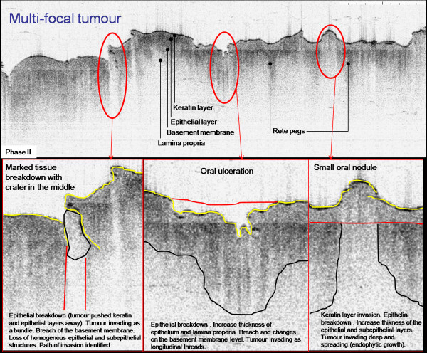
Figure 6.
An inverted OCT image of the lateral border of the tongue. There are processing artefacts running across the image. The surface differentiation is evident as visible tongue papillae poorly. 'Rete Ridges/pegs' can be seen projecting into the underlying mucosa. The various forms of tongue papillae are also visible. Histologically this area was found to represent multifocal squamous cell carcinoma; (Courtesy of Drs W Jerjes and Z Hamdoon, University College London, London)

