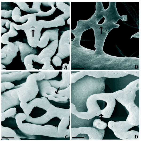
Figure 3.
Representative micrograph of different morphological pillar features in the LPerOF observed at SEM of VCC. A) and B) show pillars in the centre of the vessel or shortly distant from the bifurcation, respectively (intussusceptive branching remodelling (IBR). C) show long parallel row of pillars in the longitudinal folds of the endothelium (intussusceptive arborisation; IAR). D) show pillar formation within the capillary bed (intussusceptive microvascular growth; IMG). Black arrows indicate pillars. Bar = 10 μm.

