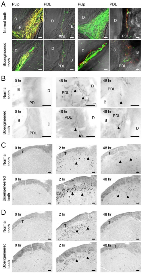
Fig. 4.
Pain response to mechanical stress. (A) Nerve fibers in the pulp and PDL in the normal (Upper) and bioengineered (Lower) tooth were analyzed immunohistochemically using specific antibodies for the combination (left 2 columns) of NF (green) and NPY (red) and the combination (right 2 columns) of NF and CGRP (red). (Scale bar, 25 μm.) (B) Analysis of galanin immunoreactivity in the PDL of a normal (Upper) and bioengineered (Lower) tooth for the assessment of orthodontic force. No galanin expression was evident in the untreated tooth (Left). Galanin expression (arrowhead) was detected in the PDL of a normal and bioengineered tooth after 48 h of orthodontic treatment (Right). (Scale bar, 25 μm.) (C) Analysis of c-Fos-immunoreactivity in the medullary dorsal horn of mice with a normal tooth (Upper) or a bioengineered tooth (Lower) after 0 h (Left), 2 h (Center), and 48 h (Right) of orthodontic treatment. c-Fos expression (arrowhead) was also detected. (Scale bar, 50 μm.) (D) Analysis of c-Fos immunoreactivity in the medullary dorsal horn of mice with a normal tooth (Upper) or a bioengineered tooth (Lower) after 0 h (Left), 2 h (Center), and 48 h (Right) of stimulation by pulp exposure. c-Fos expression (arrowhead) was evident in the medullary dorsal horn after 2 and 48 h of pulp exposure. (Scale bar, 50 μm.) D, dentin; P, pulp; B, bone; PDL, periodontal ligament; T, spinal trigeminal tract.

