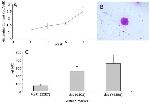
Figure 1. Characterization of Cultured Human MC.
Panel A. Histamine Content. Aliquots of CD34+ derived MC were harvested weekly and examined for average total cell histamine content. (n=14). MC histamine content increased as expected, indicating that the MC were maturing and incorporating increasing histamine content on a per cell basis. Panel B. Morphology of Cultured MCs. MC (6 weeks) were stained with Wright-Giemsa (1000 X) (1 donor representative of 13). Panel C. Cell Surface Marker Expression on MCs after 6 weeks of Culture. Surface expression of FcεRIα (22e7, n = 12) and c-kit (95C3, n= 4; YB5B8, n = 7) was determined by flow cytometry. No differences were seen between CIU subgroups and normal MC.

