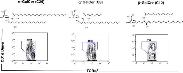
Figure 1.
Chemical structures of α and β-galactosylceramide and staining of liver NKT cells after loading of galactosylceramides onto CD1d dimers. The structures include α-GalCer (C26) with a 26 carbon acyl chain, α-GalCer (C8) with an 8 carbon acyl chain, and β-GalCer with a 12 carbon acyl chain. The phytosphingosine chains of the α-anomers differ from the sphingosine chain of the β-anomer by the presence of a second hydroxyl group. Below each structure is the two color flow cytometric pattern of representative stainings of NZB/W liver mononuclear cells with CD1d dimers loaded with each galactosylceramide versus staining for TCRαβ. Mononuclear cells were gated first on the TCRαβ+ cells. The CD1d dimer+TCRαβ+ cells are enclosed in the boxes, and the percentage of TCRαβ+ T cells within the boxes are shown.

