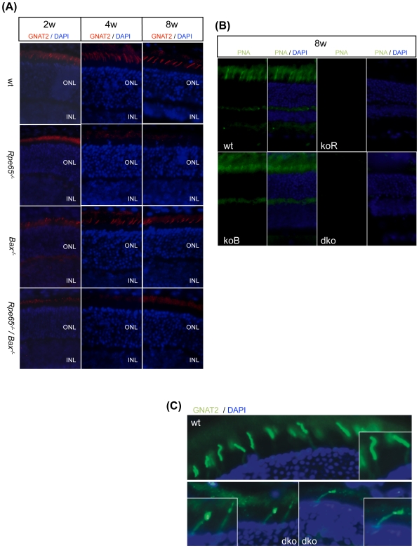
Figure 8. Early Degeneration of Cone Photoreceptors Was Not Prevented in Rpe65−/−/Bax−/− Mice.
(A) Immunohistological staining (600×) with gnat2 antibody showing that early lost expression of cone transducin, as early as 2 weeks of age, was not prevented in Rpe65−/−/Bax−/− retinas as compared to Rpe65−/− retinas (2–8 w, 2–8 weeks; ONL, outer nuclear layer; INL, inner nuclear layer). (B) FITC-labelled PNA staining (600×) in 8 week-old (8 w) Rpe65-deficient retinas confirmed that degenerating cones were not rescued in the absence of Bax. (C) Immunohistological analysis demonstrating that cone transducin failed to properly traffic to the OS in the few surviving cones still present at the retinal periphery at 6 months of age in Rpe65−/−/Bax−/− mice as compared to wt mice. Counterstaining with DAPI allowed for the identification of photoreceptor cell nuclei. koR, Rpe65−/−; koB, Bax−/−; dko, Rpe65−/−/Bax−/−.

