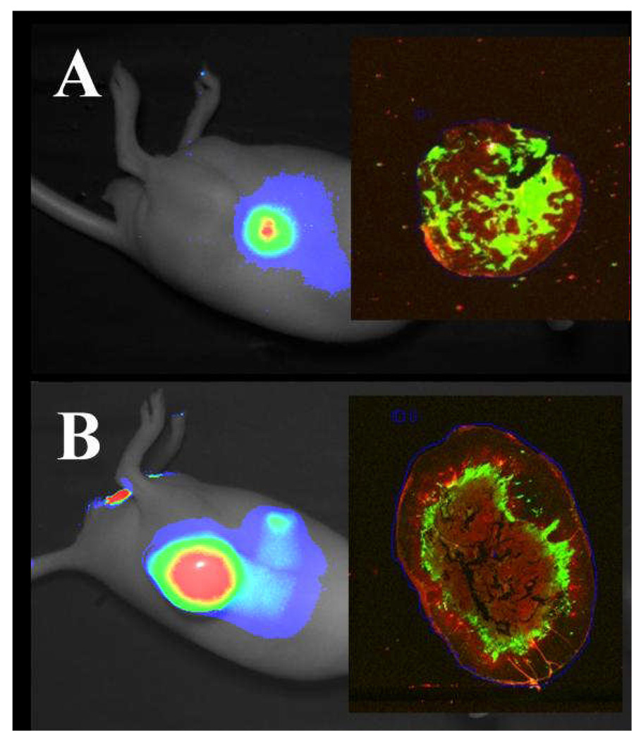
Figure 9.
Distribution of IRDye 800CW 2-DG within tumor sections. Athymic nude mice bearing 22Rv1 (A) or A431 (B) tumors were injected via the tail vein with 10 nmol or 14 nmol of IRDye 800CW 2-DG, respectively, and imaged 24 h post-injection. Excised tumors were fixed, paraffin-embedded, sectioned and scanned on the Odyssey Imaging System. Specific fluorescence in the 800 nm channel is in green and autofluorescence in the 700 nm channel is in red (inset panels).

