
Figure 6.
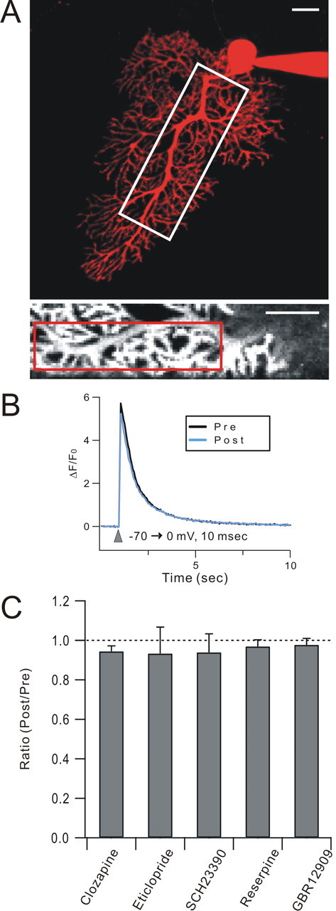
Depolarization-evoked Ca transients are not affected by DISC-attenuating drugs. A, Purkinje cells were loaded with both Fluo-5F, a Ca indicator, and Alexa Fluor 594 hydrazide, a cytosolic marker, and laser-scanning confocal microscopy was used to measure depolarization-evoked Ca transients in Purkinje cell dendrites. The top panel shows a projected z-stack confocal image of a Purkinje cell filled with Alexa Fluor 594. The bottom panel corresponds to the white box in the top panel and shows the Fluo-5F signal (at the Ca transient peak). The red box within the inset shows the region of interest used for Ca transient measurement. Scale bars: 20 μm for both top and bottom panels. B, Exemplar single unaveraged Ca traces. The depolarizing step (−70→0 mV for 10 ms) was the same as that used for DISC induction. Ca transients (measured as ΔF/F0) were plotted before (black; Pre) and after (blue; Post) the application of drugs. C, A representative set of bath-applied drugs was chosen for the screen and was applied for 20 min to mimic their use in DISC experiments. Clozapine (100 μm), eticlopride (50 μm), SCH23390 (50 μm), reserpine (30 μm), and GBR 12909 (20 μm) were used. Note that these are the higher, nonselective doses of eticlopride and SCH23390. N = 5 cells/group.