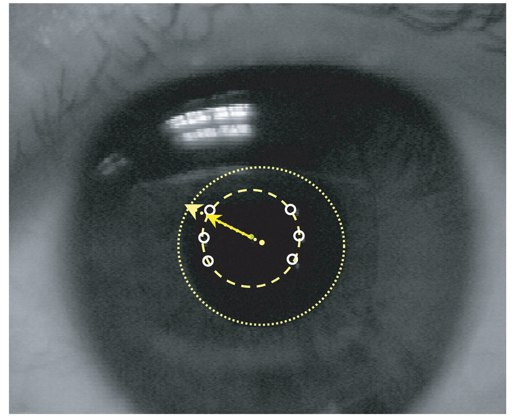
Figure B1.
An example of the image used to align subjects during data collection. The six 1st Purkinje images have been outlined and connected with the dashed circle. The pupil has been outlined using the dotted circle. Infant data were collected only when all Purkinje images fell inside the pupil. The misalignment of the instrument axis with the pupillary axis was estimated using the locations of the centers of the Purkinje image and pupil circles.

