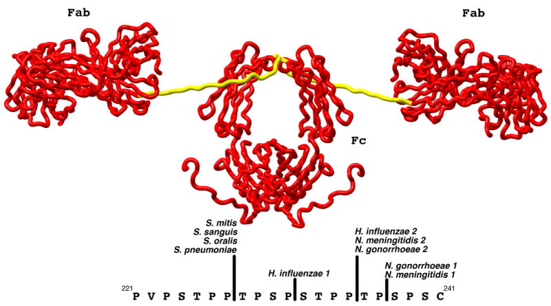
Figure 1.

Model of the structure of human IgA1 (PDB:1IGA) as determined from x-ray and neutron solution scattering and homology modeling (45). The Fab and Fc domains are rendered as red tubes and labeled while the hinge domain is rendered in yellow. The sequence of the hinge peptide and the location of the cleavage sites by various members of the IgA protease family are shown in the lower portion of the figure. The numbering utilized for the sequence of IgA1 is that of Torano and Putnam (76).
