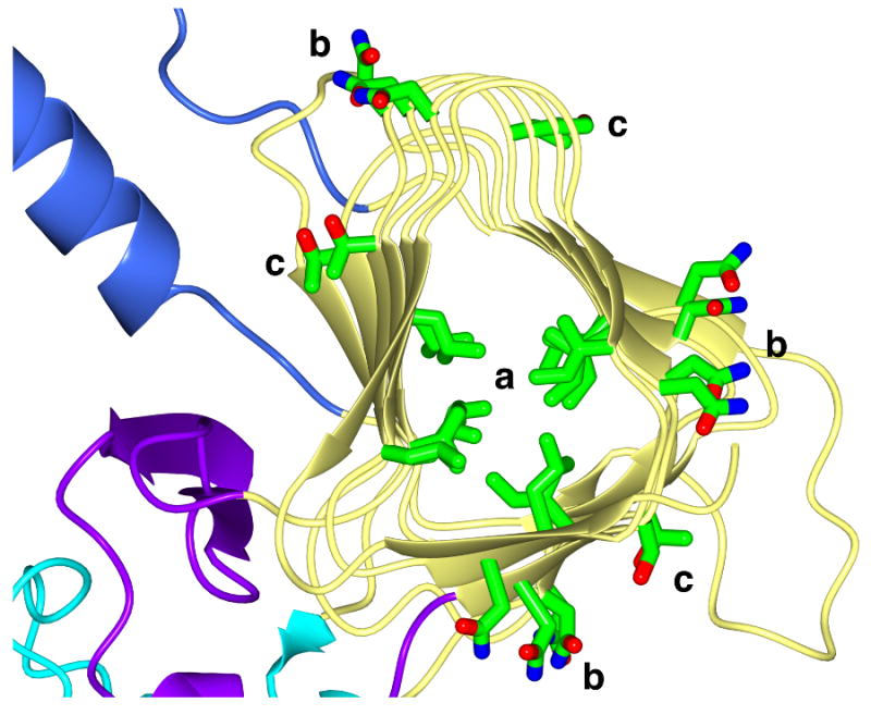
Figure 3.

Polar and hydrophobic residue stacking in the β-helix domain of IgAP. Representative stacking interactions of Ile and Leu residues (a) in the hydrophobic core as well as Asn (b) and Ser/Thr (c) stacking on the surface of the β-helix is shown. For clarity, only the region of the β-helix from residues 562-864 is depicted. The coloring of the molecule is identical to that in Figure 2.
