
Figure 7.
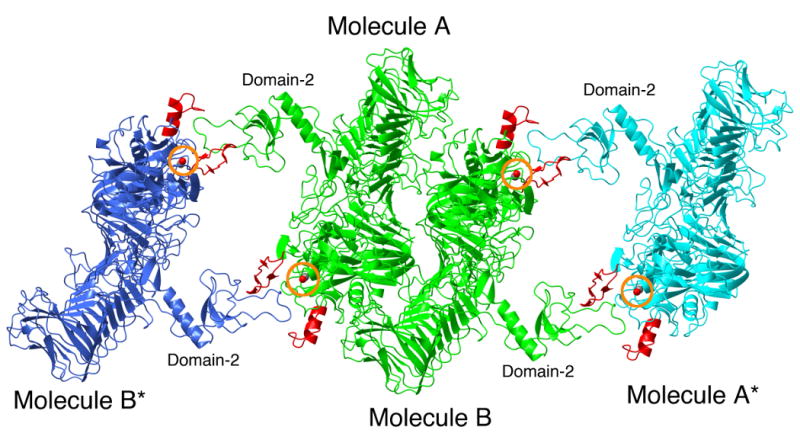
Crystal packing interactions in the structure of IgAP. The crystallographic dimer (NCS related molecules A and B) present in the asymmetric unit are rendered in green and labeled. Molecules A* and B* are related to molecules A and B by crystallographic symmetry and are rendered in cyan and blue, respectively. Loops C and D of the protease domains of all molecules are rendered in red. For the purpose of orientation, the side chain oxygen atom of the active site serine residue is visible as a red ball and circled in orange.