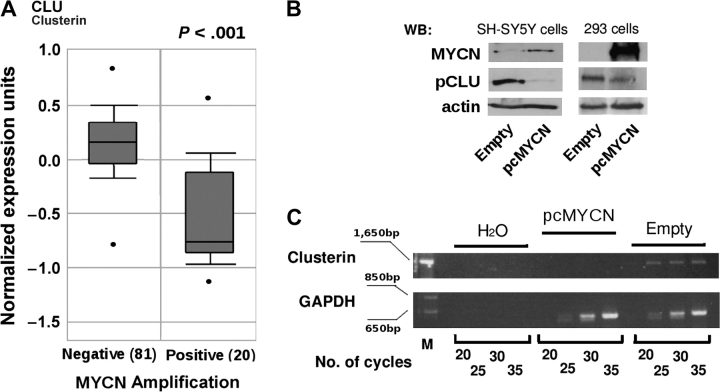
Figure 1.
Expression of clusterin and MYCN in neuroblastoma. A) Box plot of the expression of clusterin mRNA in primary human neuroblastomas with MYCN amplification (Positive) or without MYCN amplification (Negative). The figure was generated with tools found in the Oncomine Web site (www.oncomine.org), where the information regarding the neuroblastoma samples can also be found. Statistical significance was assessed by Student t test. In the Oncomine Web site, all mRNA datasets are normalized by being log2-transformed, with the median set to 0 and SD set to 1. All statistical tests were two-sided. B) WB analysis. Whole-cell lysates were prepared from SH-SY5Y and HEK-293 cells that were stably (SH-SY5Y cells) or transiently (HEK-293 cells) transfected with an MYCN (pcMYCN) or control (Empty) vector and subjected to western blot analysis with antibodies specific for MYCN, the precursor form of clusterin (pCLU), or actin (as a loading control). C) Semiquantitative reverse transcription–PCR. Clusterin mRNA expression was assessed in SH-SY5Y cells stably transfected with MYCN (pcMYCN) or with control (Empty) vector. H2O indicates PCR amplification without reverse transcription (negative control). Samples were analyzed after completion of the indicated number of PCR cycles. Amplification of glyceraldehyde-3-phosphate dehydrogenase served as a loading control. M indicates molecular weight marker. PCR = polymerase chain reaction; WB = Western blot.

