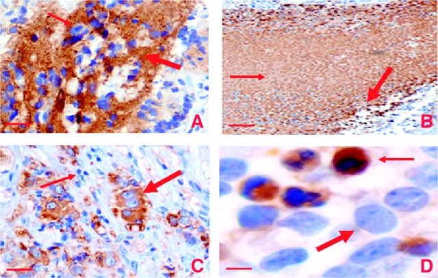
Figure 5.
Immunohistochemical staining for clusterin expression in sections from neuroblastoma tumors. Representative histological sections stained with hematoxylin and anti-clusterin (CLU) antibodies (brown color). A) Extracellular or cytoplasmic staining for clusterin. Clusterin staining in the neuropil = arrows. Original magnification was ×200. Scale bar = 0.05 mm. B) Necrotic tissue. Thin arrow = clusterin staining in necrotic tissue; thick arrow = nuclear clusterin staining. Original magnification was ×100. Scale bar = 0.1 mm. C) Ganglion cells. Thick arrow = differentiated ganglion cells; thin arrow = immature neuroblasts. Original magnification was ×200. Scale bar = 0.05 mm. D) Apoptotic cells. Thick arrow = normal nucleus; thin arrow = pyknotic nucleus. Note that only cells with fragmented or pyknotic, but not normal, nuclei are stained by the antibody against clusterin. Original magnification was ×400. Scale bar = 0.001 mm.

