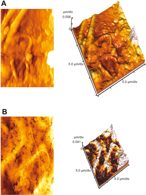
Figure 6.
Atomic force microscopy (AFM) images of rat sperm. A) 2D and 3D images of untreated cells showing cluster cells with smooth surface. B) 2D and 3D images of Fe3O4–Cu–SMA–DMSO (Smart RISUG) treated cells showing blabs in plasma membrane, disintegration leading to enzyme leaching, shortened height, and curved tail.

