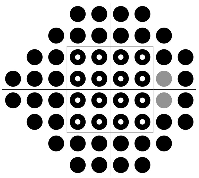
FIGURE 1.

The locations in the visual field tested in this study, as shown in the format for a right eye. The gray locations correspond to the location of the blind spot and were excluded from analysis. The gray box at (±12°, ±12°) separates the field into Central (16 locations, hollow circles) and Peripheral (36 locations, excluding the blind spot, solid circles) zones.
