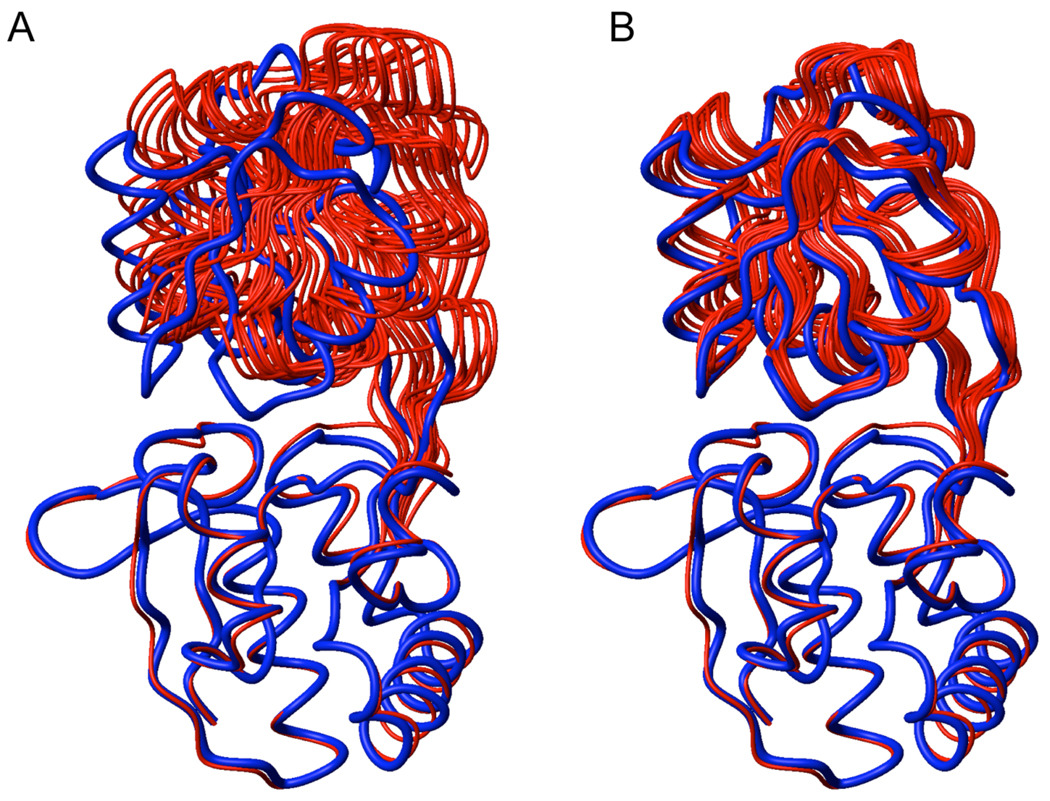
Figure 3.
PRE-based structures of bound GlnBP (red) superimposed to the X-ray model (blue; PDB ID: 1WDN) via the large domain (bottom). (A) Structures calculated by simultaneous optimization of probe conformers and protein backbone. (B) Structures calculated with previously optimized paramagnetic probe conformers using intra-domain PRE data on the fixed, open backbone coordinates. The 10 lowest-PRE energy structures (out of 200) are shown.

