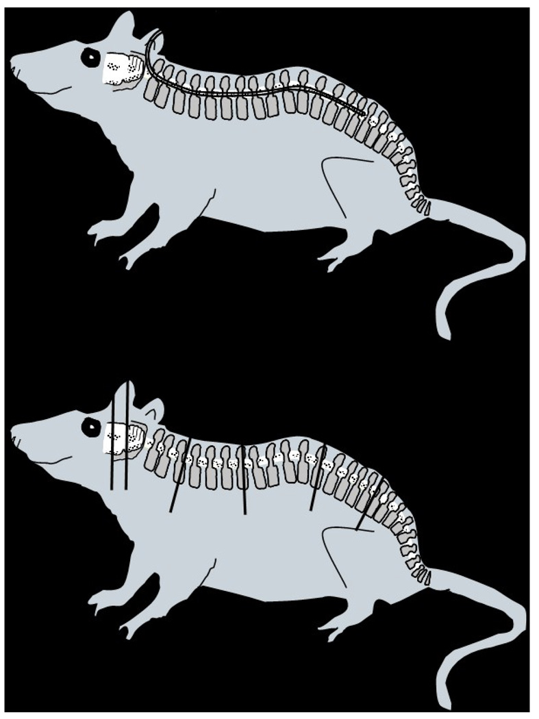
Figure 1.
(Top) An intrathecal catheter was surgically implanted in athymic female rats to permit intrathecal tumor cell inoculation and development of carcinomatous meningitis as well as intrathecal treatment administration. The PE-10 14-cm long catheter was inserted into the subarachnoid space and passed along the posterior aspect of the spinal cord to reach the lumbar area. (Bottom) In order to perform a histopathological analysis of the neuroaxis, animals were necropsied and sections from the brain, at the level of the bregma (A) and lambda (B), and the spine at the cervical (C), thoracic (D), lumbar (E) and sacral (F) levels were obtained; tissues, previously fixed in 10% formalin, were embedded in paraffin, sectioned and stained with H&E.

