Fig. 5.
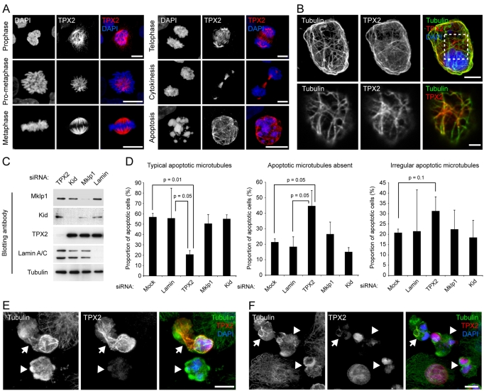
TPX2 is released from the nucleus during apoptosis, co-localises with apoptotic microtubules and is required for their efficient assembly. (A) TPX2 distribution in mitotic and apoptotic HeLa cells. TPX2 associates with spindle microtubules during mitosis, and decorates filamentous structures in apoptotic cells. (B) TPX2 co-localises with microtubules in apoptotic cells. Top panel: confocal maximum projection of an apoptotic HeLa cell (anisomycin treated) co-labelled for TPX2 and tubulin. Bottom panel: zoomed view of a single confocal section of the cortical region of interest as indicated by the hashed box in the panel above. (C,D) Silencing TPX2 disrupts apoptotic microtubule assembly. (C) Immunoblots of extracts of HeLa cells treated with siRNA oligonucleotides for 72 hours. Example shown is an experiment using oligonucleotide 1 at 50 pmol for TPX2; oligonucleotide 1 at 50 pmol for Mklp1; oligonucleotide 1 at 150 pmol for Kid; lamin at 40 pmol (refer to Materials and Methods for nomenclature). (D) Analysis of apoptotic microtubule assembly following silencing of Ran-coordinated spindle-assembly factors using the siRNA parameters described in part (C). Anisomycin-treated apoptotic cells were identified by chromatin morphology and were scored for the presence of `typical', `irregular' or `absent' apoptotic microtubules (mean + s.d.). Representative images of TPX2-silenced cells are shown in supplementary material Fig. S2B. (E,F) Examples of apoptotic microtubule arrays and co-incident TPX2 labelling in HeLa cells. (E) An apoptotic cell with strong microtubule TPX2 labelling (arrow), and an apoptotic cell lacking TPX2. In the former, microtubules are abundant, whereas in the latter, few, short microtubules can be seen. (F) An apoptotic cell with weak microtubule TPX2 staining (arrow), and apoptotic cells with TPX2 retained within the nucleus (arrowheads). In each case, microtubules are relatively sparse and poorly organised. Bars, 5 μm (A,E,F), 3 μm (B, top panel), 1 μm (B, bottom panel).

