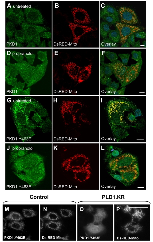
Fig. 10.
PLD1 regulates the localization of Y463-phosphorylated PKD1 to the mitochondria. (A-L) Cells were transfected with HA-tagged PKD1 or PKD1.Y463E mutant and pDsRED2-Mito (mitochondrial marker). Twenty-four hours after transfection, the cells were seeded on glass coverslips. Cells were either left untreated (A-C,G-I) or stimulated with propranolol (250 μM, 30 minutes) to block DAG formation from PA (D-F,J-L). Immunofluorescence samples were stained and processed as described in the Materials and Methods. Samples were analyzed by confocal microscopy. Green, PKD1 stained with α-P8 (α-PKD1 antibody); red, pDsRED2-Mito (mitochondria); blue, DAPI (nuclei). (M-P) Cells were seeded on eight-well μ-slides and transfected with GFP-tagged PKD1.Y463E, pDsRED2-Mito (mitochondrial marker) and control vector or PLD1.KR. Twenty-four hours after transfection, the cells were fixed and analyzed. The experiments were performed three times and similar results were obtained.

