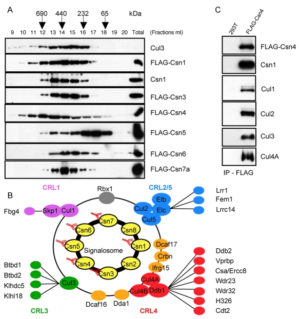
Fig. 1.
The protein interaction network of the mammalian CSN. (A) Protein extracts prepared from HEK 293T cells stably expressing the indicated FLAG-tagged CSN subunits were fractionated on a Superose 6 column (bed volume: 24 ml) and 1-ml fractions were collected and analyzed by SDS-PAGE with specific FLAG, Csn1 and Cul3 antibodies. (B) CSN-interacting CRL subunits are highlighted in purple (SCF), blue (CRL2 and CRL5), green (CRL3) and red (CRL4). Subunits highlighted in orange have been previously found associated with CRL4s. (C) CSN-cullin interactions observed by LC-MS-MS were confirmed by co-immunoprecipitation experiments combined with western blot identification.

