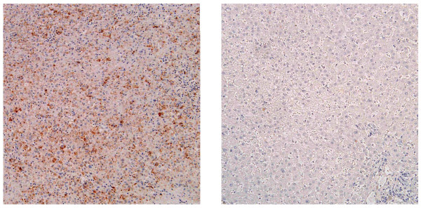
Figure 1.
Twist expression in HCC tissue and its adjacent liver tissue. Twist expression was assessed by immunohistochemical staining in a human HCC tissue (left panel) and corresponding tumor-free section (right panel). Photographs were taken using a light microscope (BX51, Olympus) equipped with a digital image capturing system (DP50, Olympus). Shown are the representative images of twist expressing a HCC tissue and its corresponding tumor-free section.

