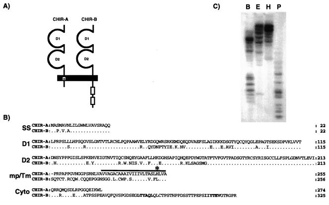
Figure 1.
(A) Schematic representation of the predicted CHIR-A and CHIR-B molecules with two Ig-like extracellular domains. CHIR-A encodes a short cytoplasmic region and a transmembrane segment with a charged histidine (H) residue. CHIR-B possesses a long cytoplasmic tail with two ITIM units (outline boxes) and a nonpolar transmembrane region. (B) Comparison of the CHIR-A and CHIR-B amino acid sequences. In this alignment, the relatives are numbered with reference to the start of the signal sequence. Conserved residues in CHIR-B are represented as dots. The solid bar indicates the putative transmembrane region; an asterisk marks the positively charged histidine residue of CHIR-A; and the ITIM-units of CHIR-B are highlighted in bold. (C) Southern blot analysis of the Chir gene family. DNA from nucleated chicken erythrocytes was digested with BamHI (B), EcoRI (E), HindIII (H), and PstI (P) and analyzed with a probe corresponding to the extracellular region of CHIR-B. Accession nos: CHIR-A, AF306851; CHIR-B, AF306852.

