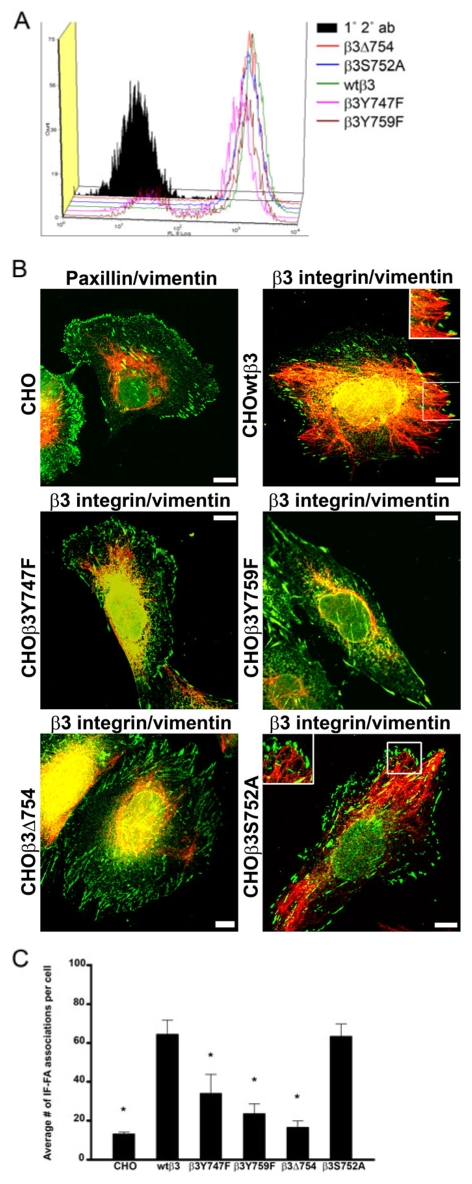
Fig. 3.
IF recruitment to FAs is regulated by β3 integrin in CHO cells and specifically by tyrosine residues within the tail of the β3 integrin. (A) Constructs encoding wild-type and mutant GFP-tagged β3 integrin were transfected into CHO cells and surface expression of αvβ3 integrin was evaluated by FACS using LM609 antibody (1° 2° ab, nontransfected CHO cells labeled with primary and secondary antibodies). (B) CHO cells (top left) were prepared for immunofluorescence using a combination of paxillin (green) and vimentin (red) antibodies. In addition, CHO cells expressing the indicated GFP-tagged β3 integrin proteins were stained for vimentin (red). The insets (top right, bottom right) are higher magnifications of the boxed areas. (C) Quantification of IF-FA association for all of the CHO cell lines. Over 200 cells were analyzed per cell line in at least three separate experiments. Error bars represent s.e.m. of three experiments. *P<0.03; statistically significant decrease in IF-FA association compared with CHO cells expressing wild-type β3 integrin.

