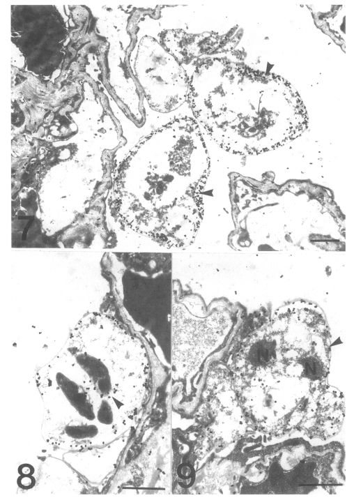
Figs. 7-9.
Fig. 7. Immunogold labelling for tubulin on a cystic form of Pneumocystis carinii. The number of gold particles is remarkably increased and densely concentrated on the inner electron-lucent layer of the cell wall of the two cystic forms. Bar = 1 µm. ×5,000. Fig. 8 Immunogold labelling for tubulin on a cystic form of Pneumocystis carinii. The gold particles are observed at the pellicle of the intracystic bodies (arrowhead). Bar = 1 µm. ×8,000. Fig. 9 Immunogold labelling for tubulin on a trophic form of Pneumocystis carinii. The gold particles are distributed on the cell wall (arrowhead) and the peripheral cytoplasm. N, nucleus. Bar = 1 µm. ×5,000.

