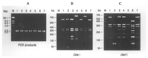
Fig. 3.
(A) Agarose gel electrophoretic pattern of PCR products of SSU rDNA of 7 types of Acanthamoeba isolates; M, Hind III digested λ phage DNA. (B)(C) Electrophoretic patterns of restriction fragments of PCR product from 7 types of Acanthamoeba isolates by Dde I and Hinf I; B, Agarose gel; C, Polyacrylamide gel; M, 50 bp ladder (BM, Germany); Lane 1, KA/LS1; 2, KA/LS2; 3, KA/LS5; 4, KA/LS4; 5, KA/LS18; 6. KA/LS7; 7, KA/LS31.

