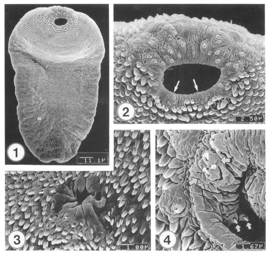
Figs. 1-4.
Scanning electron microscopic (SEM) view of metacercariae of Metagonimus takahashii. Fig. 1. Ventral view of an excysted metacercaria. Fig. 2. Tegument around the oral sucker. Note distribution of seven type II sensory papillae (arrows) on the lip of the oral sucker. Fig. 3. Tegument around the ventral sucker showing dense distribution of tegumental spines. Fig. 4. Inner side of the lip of the oral sucker showing small and large type I sensory papillae (arrows).

