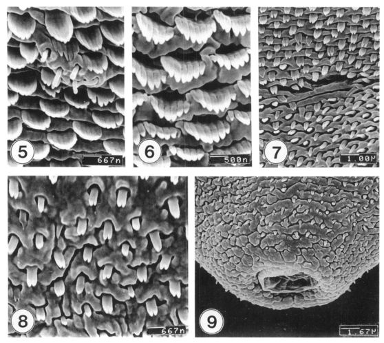
Figs. 5-9.
SEM view of metacercariae of M. takahashii. Fig. 5. Tegumental spines and type I sensory papillae at ventral surface between oral and ventral suckers. Fig. 6. Spines at antero-dorsal surface having 7-8 pointed tips. Fig. 7. Dorsal tegument near the Laurer's canal. Fig. 8. Ventral tegument anterior to the excretory pore showing peg-like spines with two-pointed tips. Fig. 9. Tegument around the excretory pore. Spines are sparse, and cytoplasmic processes are cobblestone-like.

