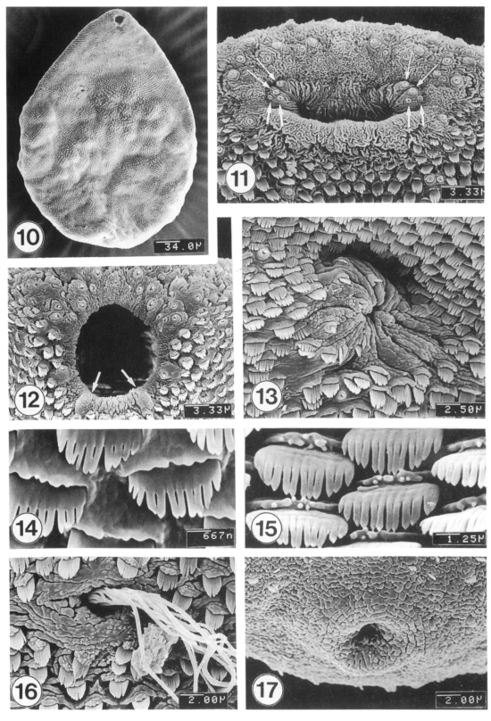
Figs. 10-17.
SEM view of 1-4-week-old adults of M. takahashii. Fig. 10. Ventral view of a whole worm, 2-week-old. Fig. 11. Oral sucker typically showing two pairs each of large (thin arrows) and small type I papillae (thick arrows; other two pairs are hidden at ventrolateral sides) inside the oral sucker. Fig. 12. Tegument around the oral sucker showing type I and type II (seven in number; arrows) sensory papillae on the lip. Fig. 13. Tegument around the ventral sucker showing densely distributed spines. The ventral sucker is muscular and showing many wrinkles. Fig. 14. Ventral tegument between oral and ventral suckers showing broad, brush-like tegumental spines with 9-12 pointed tips. Fig. 15. Mid-dorsal tegument showing brush-like spines with 10-13 tips. Fig. 16. Dorsal tegument around the Laurer's canal. Many sperms are seen entering into the canal. Fig. 17. Tegument around the excretory pore showing very few spines.

