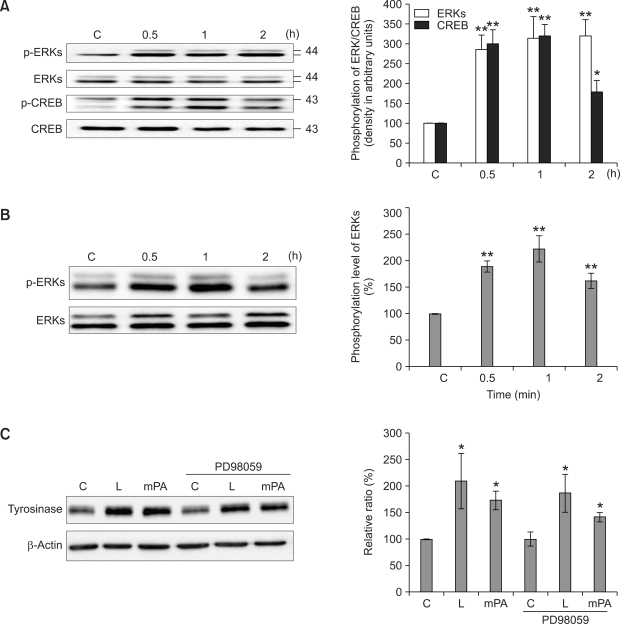
Figure 3.
Signaling pathways activated by lotus flower essential oil in human melanocytes. Supplement-reduced melanocytes were treated with 10 µg/ml of lotus flower essential oil (A) or 50 µM of palmitic acid methyl ester (mPA) (B) for 0.5, 1, or 2 h. The whole lysates were electrophoresed via SDS-PAGE and analyzed by immunoblotting with each antibody. The intensity of phosphorylation and total ERKs and CREB bands were quantitated by densitometry and the amounts of phosphorylated ERKs and CREB were normalized versus total ERKs and CREB. The data represent the means ± S.E. of four independent experiments. Supplement-reduced melanocytes were treated with 10 µg/ml of lotus flower essential oil (L) or 50 µM of palmitic acid methyl ester (mPA) in the absence or presence of 10 βM PD98059 (PD98059) for 2 days. The whole lysates were electrophoresed via SDS-PAGE and analyzed by immunoblotting with each antibody. Tyrosinase expression was normalized versus β-actin. The data represent the means ± S.E. of three independent experiments C: unstimulated melanocytes. *P < 0.01; **P < 0.001.

