
Figure 3. GO-201 induces S phase arrest and necrosis.
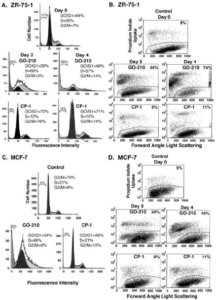
A-D. ZR-75-1 (A and B) and MCF-7 (C and D) cells were treated with 5 μM GO-201 or CP-1 each day for 3 and 4 d. Cells were fixed and analyzed for cell cycle distribution by flow cytometry (A and C). The percentage of diploid cells in G0/G1, S and G2/M phases is included in the panels. There were no distinct sub-G1 peaks supporting induction of apoptosis; whereas, debris in this region reflects cells with DNA degradation and is included as a percentage of diploid cells. Cells were also stained with propidium iodide and analyzed by flow cytometry for necrosis (B and D). The percentage of necrotic cells is included in the panels.