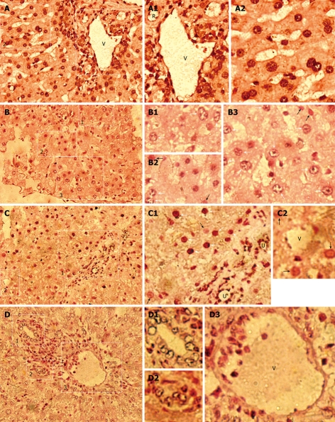
Figure 2.
Human donor contributions in HRC liver analyzed by in situ hybridization (ISH) for human Alu sequences. Black/dark brown Alu-positive human cells in nuclei (white arrow) were identified in receipt liver using a probe specific for human DNA Alu sequences, and counterstained (pink) with nuclear fast red. Black/dark brown hybridization signals (white arrow) shows Alu-positive human cells in liver of MTR and human samples, whereas no Alu-positive human cells were found in normal control (NC) rats (data not shown). White arrow and black arrow indicate the partially demonstrated Alu-positive and negative human cells in B1-B3, C1 and C2, respectively. A: Human liver (positive control); B-D: Detection of donor-derived human cells in the chimeric liver of HRC. R: Artery; U: Bile duct; V: Vein. A: Low-power magnification of hAlu-positive human cells; A1: High-power magnification of the region (1) in the right rectangle of (A) demonstrating hAlu-positive vein (V) containing hAlu-positive human endothelial cell-like cells (hECLs), hAlu-positive artery (R) containing hAlu-positive human cells, and hAlu-positive human hepatocytes and other human cells in human liver; A2: High-power magnification of the region (2) in the left rectangle of (A), hAlu-positive human hepatocytes and other human cells in human liver; B: Low-power magnification of hAlu-positive human cells; B1: High-power magnification of hAlu-positive human cells (white arrow) of the rectangle region (1) in (B); B2: High-power magnification of hAlu-positive human cells (white arrow) and hAlu-negative cells (white arrowhead) of the rectangle region (2) in (B); B3: High-power magnification of hAlu-positive human cells (white arrow) and hAlu-negative cells (white arrowhead) of the rectangle region (3) in (B); C: Low-power magnification of hAlu-positive human cells; C1: High-power magnification of the rectangle region (1) in (C) showing hAlu-positive human hepatocyte-like cells (hHLCs) (white arrow), hAlu-positive bile duct (U) epithelial cells and other human cells (white arrow) in chimeric liver; C2: High-power magnification of the rectangle region (2) in (C) showing hAlu-positive hECLs in vein (V) of chimeric liver; D: Low-power magnification of hAlu-positive small bile ducts (U) and hAlu-positive vein (V) in chimeric liver; D1, D2: High-power magnification of hAlu-positive small bile ducts (U) in (1, 2) of (D), respectively; D3: High-power magnification of vein (V) containing hAlu-positive hECLs in (3) of (D).

