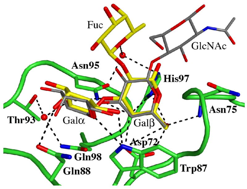
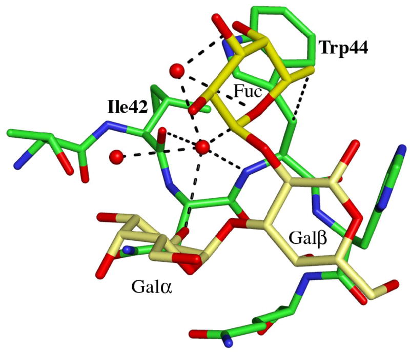
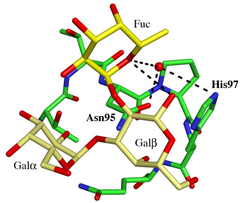
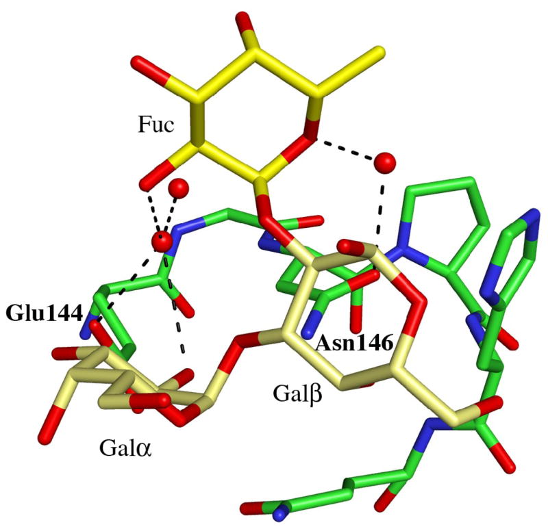
Figure 2.
Details of the blood group B binding sites. (a) The blood group B trisaccharide (yellow) bound to the β-site. Direct and indirect hydrogen bonds between the ligand and the protein are indicated by dashed lines (cut-off at 3.4 Å). For comparison, the xenotransplantation epitope trisaccharide (gray), PDBid 2IHO, is superimposed onto the binding site. (b)–(d) The interactions provided by the fucose residue are indicated by dashed lines, hydrogen bonds with a cut-off at 3.4 Å and C-C interactions with a cut-off at 4.0 Å for (b) the α-site, (c) the β-site and (d) the γ-site, respectively. Water molecules are represented as red spheres.


