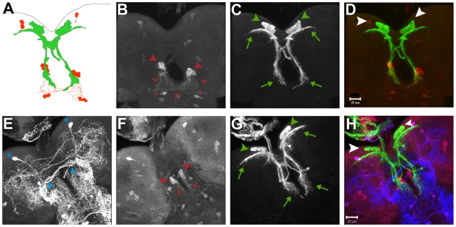
Figure 4. Ddc and serotonin labeled cells in larval brains do not overlap with IPCs.
(A) A schematic drawing depicting a third instar larval brain with the relative positions of the IPCs and their processes (in green) and the Ddc labeled cells and their processes (in red). The cellular processes from the two domains seem to intermingle in the sub-esophageal ganglia region. (B–H) Three-dimensional projections of confocal Z-stacks of a wild-type Drosophila larval brain from a wandering third instar larva expressing mCD8GFP with Dilp2GAL4 and immunostained with anti-serotonin antibody (E), anti-Ddc antibody (B, F) and anti-GFP antibody, (C, G). (D) is a merge of (B) and (C) while (H) is a merge of (E),(F) and (G). In (D) and (H), anti-Ddc staining is in red and anti-GFP in green while anti-serotonin is blue in (H). Red arrowheads in (B, F) indicate Ddc stained cells in the sub-esophageal ganglia that lie in close proximity to IPC projections (bottom green arrows in C, G). Smaller red arrowheads indicate cells which send out processes (marked with red arrows) that seem to intermingle with these IPC projections. Green arrowheads in (C, G) mark the IPCs in the two brain lobes. Green arrows indicate the projections of the IPCs towards the lateral protocerebrum (top green arrows) and sub-esophageal ganglion (bottom green arrows). Ddc marked cells (indicated by big red arrowheads in B, F) stain with the anti-serotonin antibody (E, marked by blue arrowheads), but have lesser serotonin staining than some neighboring cells (for example, cells in the lateral protocerebrum indicated by blue asterisk in E). Scale bars B–H 20 µm.

