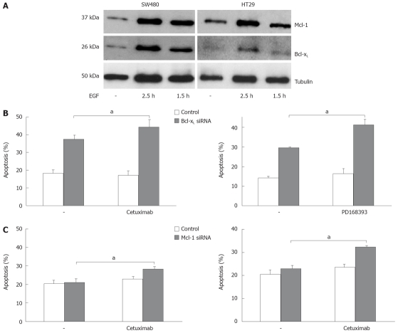
Figure 5.
Bcl-xL and Mcl-1 knock down enhances apoptosis induction after EGFR1 inhibition. A: SW480 and HT29 cells were treated with EGF (100 ng/mL) for the time indicated. Whole cell lysates were prepared, separated, and immunoblotted with antibodies against Bcl-xL, Mcl-1 and α-tubulin; B: SW480 cells were transfected with siRNA specific for Bcl-xL (upper panel), or Mcl-1 (lower panel), respectively, or transfected with siRNA specific for GFP as control. 24 h post transfection cells were treated with cetuximab (100 μg/mL) or PD168393 (0.7 μmol/L) for further 24 h. Cells were then harvested and analyzed for apoptosis induction by flow cytometry. Assays were performed in triplicates and are representative for at least two independent experiments. Values are means ± SD, aP < 0.05.

