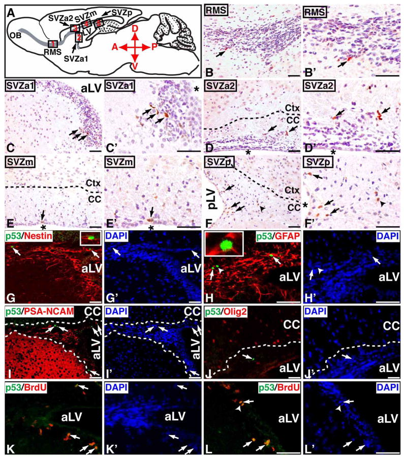
Figure 6. A minor population of SVZ stem and progenitor cells exhibit a detectable mutant p53ΔE5-6 protein expression in young adult brain.
(A) Schematic drawing of the sagittal plane of the adult mouse brain. Except for the dentate gyrus of the hippocampus, all the Nestin+ cells in the adult brain are restrictively located in the SVZ and RMS colored in grey. Different parts of the RMS and SVZ are shown in five designated boxed areas. At 2 months of age, most of the p53ΔE5-6-positive cells (arrows, B to F′) are identified in the adult neurogenic areas, which include the RMS (B, B′), the anterior SVZ (SVZa1, C, C′ and SVZa2, D, D′), medial SVZ (SVZm, E, E′), and posterior SVZ (SVZp, F, F′). The arrowhead in (F, F′) point to a p53ΔE5-6-positive cell in the adjacent corpus callosum. Sections of the anterior SVZ from 2-month-old p53ΔE5-6 mice were stained by anti-p53/anti-Nestin (G) and DAPI (G′); anti-p53/anti-GFAP (H) and DAPI (H′); anti-p53/anti-PSA-NCAM (I) and DAPI (I′); anti-p53/anti-Olig2 (J) and DAPI (J′); and anti-p53/anti-BrdU (K, L) and DAPI (K′, L′). Arrows in (G-L′) point to the p53ΔE5-6-positive cells in the SVZ. An arrowhead in (H, H′) points to a p53ΔE5-6-positive/GFAP-negative progenitor cell. The insets in (G, H) show the high-magnification views of the p53ΔE5-6-positive cells expressing Nestin and GFAP, respectively. Arrowheads in (L, L′) point to a p53ΔE5-6-positive cells during mitosis. aLV and pLV, anterior and posterior lateral ventricle; “*”, lateral ventricle. Scale bar, 25 μm.

