Figure 1.

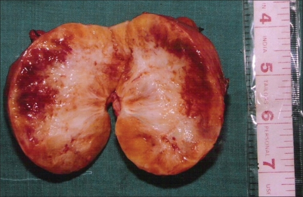
Photograph of cut section of the gross specimen of the Retroperitoneal Schwannoma showing a firm tumor with areas of haemorrhage and patchy areas of necrosis

Photograph of cut section of the gross specimen of the Retroperitoneal Schwannoma showing a firm tumor with areas of haemorrhage and patchy areas of necrosis