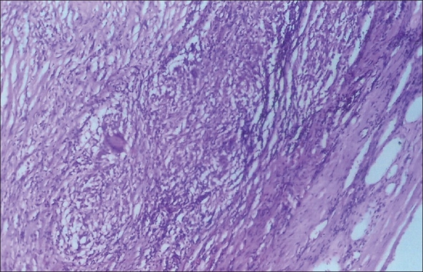
Figure 2.

Photomicrograph of the peripheral aspect of Schwannoma showing capsule, interlacing fascicles of spindly cells and epithelioid cell granulomata. Note a Langhans Giant cell. (H&E, ×100)

Photomicrograph of the peripheral aspect of Schwannoma showing capsule, interlacing fascicles of spindly cells and epithelioid cell granulomata. Note a Langhans Giant cell. (H&E, ×100)