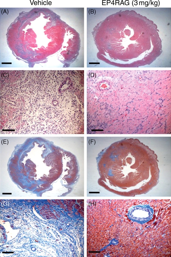
Figure 2.

HE and Masson-trichrome staining. Low-power (A, B, E, and F) and high-power (C, D, G, and H) photomicrographs of HE-stained (A through D) and Masson-trichrome-stained (E through H) LV cross-sections obtained from I/R (A, C, E, and G), I/R + EP4RAG (3 mg/kg) (B, D, F, and H) rats 7 days after operation. EP4RAG-treated I/R rats had significantly smaller LV diameters compared with those of vehicle. EP4RAG attenuated infiltration of inflammatory cells and LV fibrosis (D and H) compared with vehicle (C and G). Scale bars: 2 mm (A, B, E, and F) and 100 µm (C, D, G, and H).
