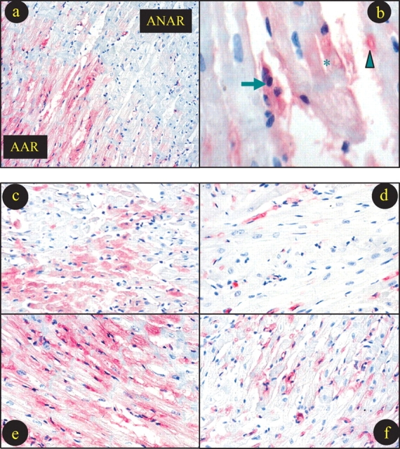
Figure 3.

Typical photographs for myeloperoxidase (MPO) immunostaining. (a) Sample from a hypercholesterolaemic rabbit treated with vehicle showing that intensive MPO staining presents in area-at-risk (AAR), but not in area-not-at-risk. (b) High-power magnification photograph showing MPO staining in leucocytes (arrow), microvessel (arrowhead) and cardiomyocyte (star). Remaining photographs (c–f) were taken from AAR with different treatments. (b) Normal diet treated with vehicle; (c) normal diet treated with rosiglitazone (RSG); (d) high-cholesterol diet treated with vehicle; (e) high-cholesterol diet treated with RSG.
