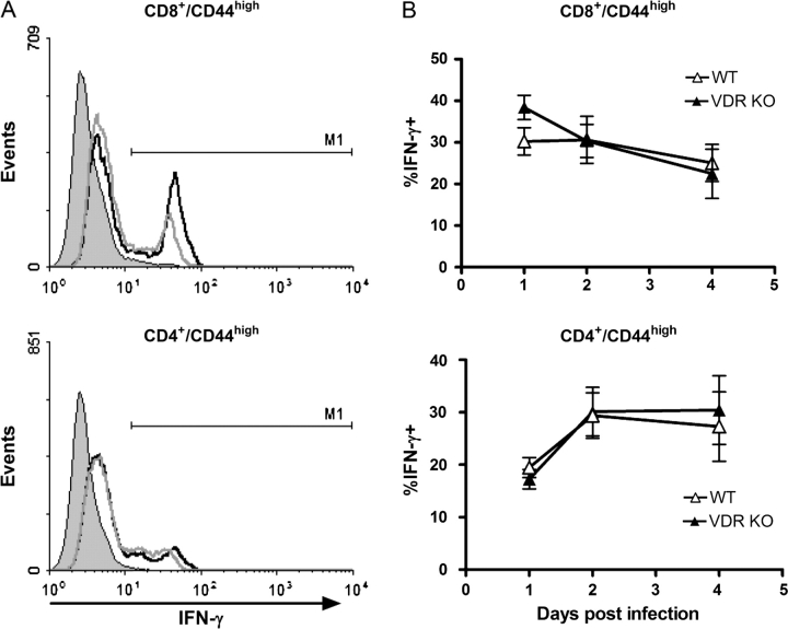
Fig. 3.
IFN-γ secretion by memory T cells in young VDR KO and WT mice after secondary exposure to Listeria monocytogenes. Splenocytes were stained for IFN-γ, CD8, CD4 and CD44 following 1–4 days post-secondary infection. (A) CD8+/CD44high (top) or CD4+/CD44high (bottom) T cells were gated and the histograms showing staining for IFN-γ-producing cells presented. The solid peak shows the isotype control staining and IFN-γ expression of VDR KO (black line) and WT (gray line). (B) Average percentage of IFN-γ-producing CD8/CD44high (top) and CD4/CD44high (lower) T cells in young VDR KO and WT mice. Values are mean ± standard error of the mean of four individual mice. Experiments were repeated twice.

