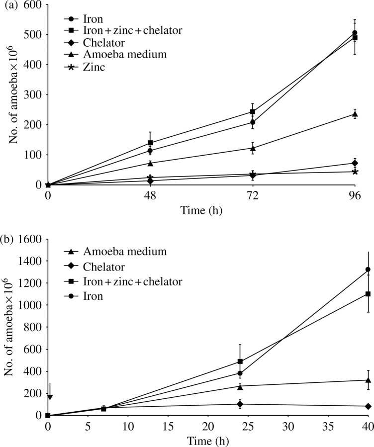
Figure 2.
(a) Inhibition of trophozoite growth by iron starvation. An initial 5×10³ HM1:1MSS E. histolytica trophozoites were inoculated per tube with TYI medium alone (filled triangles), 30 µM ferrous sulphate (filled circles), 1,10-phenanthroline (filled diamonds), 50 µM zinc sulphate (asterisks) or 30 µM ferrous sulphate+50 µM zinc sulphate+1,10-phenanthroline (filled squares), and counted at 48, 72 and 96 h. (b) Chelator-dependent inhibition is reversed by iron saturation. An initial 5×10³ HM1:1MSS E. histolytica trophozoites were inoculated per tube of TYI medium with 30 µM 1,10-phenanthroline for 7 h. After this initial exposure to the chelator, trophozoites were exposed to 30 µM ferrous sulphate (filled circles), 30 µM 1,10-phenanthroline (filled diamonds) or 30 µM ferrous sulphate+50 µM zinc sulphate+30 µM 1,10-phenanthroline (filled squares), or left in TYI medium alone (filled triangles), and counted at 7, 24 and 40 h. The time when second additions were supplemented (after the 7 h) is indicated by an arrow.

