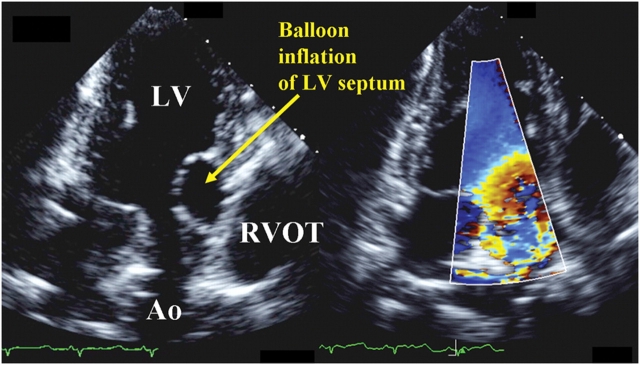
Figure 1.
Epicardial echocardiograms of a sheep at the apical three-chamber window showing balloon inflation of the left ventricular (LV) septum at the LV outflow tract (OT), mimicking clinical upper septal hypertrophy and distorting the LVOT geometry (left panel). Colour Doppler revealed non-uniform flow across the LVOT (right panel). RV, right ventricle; Ao, Aorta.

